|
|
|
Dr Neville Davis is a Queensland surgeon with vast experience in the treatment of melanoma. He wrote
The skin & melanocytes (pigment cells) Melanocytes are found distributed in the skin:
|
|
|
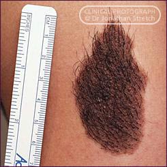
A congenital hairy naevus |
Acquired and congenital naevi (moles) Melanocytes and naevus cells do not normally aggregate together to form naevi (moles) until after the first few years of life. Occasionally children are born with pigmented birth marks (congenital naevi). However, the vast majority of naevi are acquired during the ages of 5 - 20 yrs. During this time new naevi develop and others change, but usually in some proportion to other naevi and overall body growth. The development of new naevi or change in existing ones, especially of single naevi after adolescence is significant and warrants expert assessment. |
 |
 |
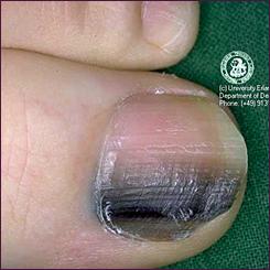
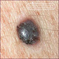
| Subungual melanoma: melanoma beneath the toe-nail | In-situ melanoma: (very superficial/early) |
 |
|
| Melanoma of the scalp | Thick Melanoma: Breslow thickness 1.7mm |
 |
 |
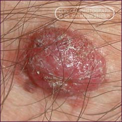
| Amelanotic melanoma: This melanoma is composed almost entirely of cells which no longer produce melanin pigment. It appears to have arisen from the lightly pigmented (tan) coloured naevus of which a remnant remains along its lower edge. |
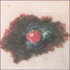
Advanced
melanoma: This melanoma is very interesting because a clone
(group) of cells within the tumour have become sufficiently abnormal
that they have lost the capacity to produce the characteristic dark
(melanin) pigment. These cells are also dividing relatively quickly
and producing the raised nodule. This has a rich blood supply and
hence the bright red colour.
|
|
Factors recognized as increasing the risk of melanoma
How does melanoma develop Like cancers in other parts of the body, melanomas are composed of cells which multiply without the normal control of the body's regulating systems. Just as the specific cause of most cancers is not fully understood, it has not yet been possible to completely identify how melanoma develops. However it has been determined that living in climates with high levels of ultraviolet light greatly increases the incidence of melanoma. It appears that the ultraviolet (ionizing radiation) may directly mutate (corrupt) segments of the genetic code located in melanocyte chromosomes and responsible for cell control. Melanoma results at least in part from the interaction of the accumulated ultraviolet irradiation from the sun and the varying ability of different skin types to resist this damage. Nonetheless melanoma does not always occur in body parts which have received the most solar exposure. It is important to understand that cancers do not develop directly from normal cells but progressively evolve in a series of stages that can frequently be readily identified with expert examination. A number of changes in the skin can be identified as representing changes in the pigment cells which are pre-cancerous.
Examination
of the skin
When are naevi (moles) excised
If naevi are stable and do not have any specific clinical features of concern, removing them does not offer any protection against the development of melanoma.
More about melanoma |
|
|
Jonathan
Stretch Plastic Surgeon D.Phil (Oxon) F.R.A.C.S.
|
|
 Characteristic
patterns of aggregation can usually be recognized by expert clinicians
as those forming junctional, dermal, compound and dysplastic naevi
(moles). The word mole is an old English description which
originally referred to dark hairy patches on the skin (congenital
naevi) which were likened to the soil burrowing animals.
The term mole has since been used much more broadly and as such
is not clinically useful. Melanoma can develop in the scattered
melanocytes in plain skin or associated with pre-existing pigmented
naevi.
Characteristic
patterns of aggregation can usually be recognized by expert clinicians
as those forming junctional, dermal, compound and dysplastic naevi
(moles). The word mole is an old English description which
originally referred to dark hairy patches on the skin (congenital
naevi) which were likened to the soil burrowing animals.
The term mole has since been used much more broadly and as such
is not clinically useful. Melanoma can develop in the scattered
melanocytes in plain skin or associated with pre-existing pigmented
naevi.